View Posterior Tibial Pulse Vs Dorsalis Pedis Pics. It arises at the anterior aspect of the ankle joint and is a continuation of the anterior tibial artery. Browse the newest dorsalis pedis and posterior tibial study sets and find the tools you need to get ahead today!
Tibial Pedal Arterial Access Retrograde Interventions For Advanced Peripheral Arterial Disease Critical Limb Ischemia from www.openaccessjournals.comA diminished foot pulse may be the only clue that a patient is at increased risk of cardiovascular death. (1) palpate at inner aspect of posterior malleolus (in the groove between the malleolus and the achilles tendon). Inspect the feet for colour, temperature and presence of edema.
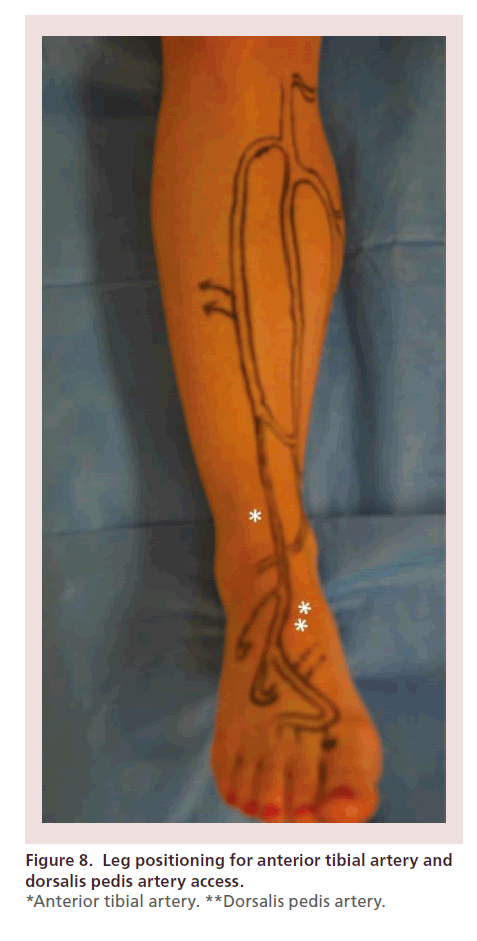
There is a faint palpable dorsalis pedal pulse, but no posterior tibial pulse.
The dorsalis pedis angiosome encompasses the entire dorsal aspect of the foot through the variable medial tarsal, lateral tarsal, arcuate and dorsal metatarsal arteries (fig. No disagreement in dp pulses. Dp = dorsalis pedis (pulse felt at top of foot), and pt = posterior tibial (pulse felt behind the medial ankle bone). Dorsalis pedis (dp) and posterior tibial (pt) pulses were palpated and were then examined by doppler with measurement of systolic pressures.


Tidak ada komentar:
Posting Komentar